


Useful articles

Liver Hydatid Cyst (Echinococcosis): Symptoms, Diagnosis and Treatment
Doctor's blog
sbdrmn
Direction: Robotic surgery · All services
Liver echinococcosis, or a hydatid cyst of the liver, is a disease in which the larva of a tapeworm carried by dogs forms a fluid-filled cyst in the liver. It may cause no symptoms for years, but as the cyst grows it brings a risk of rupture and other serious complications. Below we explain in plain language how infection happens, which symptoms should worry you, how the diagnosis is made, what treatment options exist and how to protect yourself.
Contents
- What echinococcosis is and how people catch it
- Symptoms of a liver hydatid cyst
- Complications: why a cyst cannot simply be ignored
- Diagnosis: ultrasound, CT and blood tests
- Treatment options for liver echinococcosis
- Follow-up after treatment and recurrence
- How to prevent echinococcosis
- Frequently asked questions
What echinococcosis is and how people catch it
Echinococcosis is a disease caused by the larva of a small tapeworm, Echinococcus granulosus. The adult worm lives in the intestine of dogs (and of wolves and jackals), and its eggs pass out in the faeces onto soil, grass and into water. When a sheep, a cow or a person swallows these eggs, the larva passes through the bowel wall into the bloodstream and most often settles in the liver, less often in the lungs, where it forms a fluid-filled bladder — a hydatid cyst.
The main ways people become infected are:
- close contact with dogs — stroking, kissing, sharing dishes; the eggs stick to the dog's coat;
- unwashed vegetables, herbs and fruit — especially from a garden where dogs roam;
- unboiled water from irrigation channels, springs and other open sources;
- working with livestock — herding and slaughtering sheep; the cycle closes when dogs are fed raw offal.
In Uzbekistan, where livestock farming is widespread and many households keep dogs in the yard, echinococcosis remains a real public health problem. The disease is not passed from person to person.
Symptoms of a liver hydatid cyst
The cyst grows very slowly and often causes no symptoms at all for years. It is frequently found by chance on an ultrasound done for another reason. Once the cyst becomes large, the following may appear:
- heaviness, a feeling of fullness or a dull ache under the right ribs;
- a bulge in the upper abdomen or an enlarged liver;
- loss of appetite, nausea, feeling full quickly;
- allergic rashes and itching.
When the lungs are affected, cough, chest pain or breathlessness may occur — these are investigated separately.
Complications: why a cyst cannot simply be ignored
A large cyst presses on neighbouring organs, and the risk of complications grows over time: infection and abscess formation, rupture into the bile ducts with jaundice, or rupture into the abdominal or chest cavity. When a cyst ruptures, its fluid can trigger a severe allergic reaction — anaphylaxis — and seed new cysts.
Call an ambulance immediately (103) if a person with a known liver cyst suddenly develops severe abdominal pain, breathlessness, a rash all over the body, a drop in blood pressure with weakness, high fever with shivering, or yellowing of the eyes and skin. Protect the abdomen from blows and heavy lifting — these can provoke rupture of the cyst.
Diagnosis: ultrasound, CT and blood tests
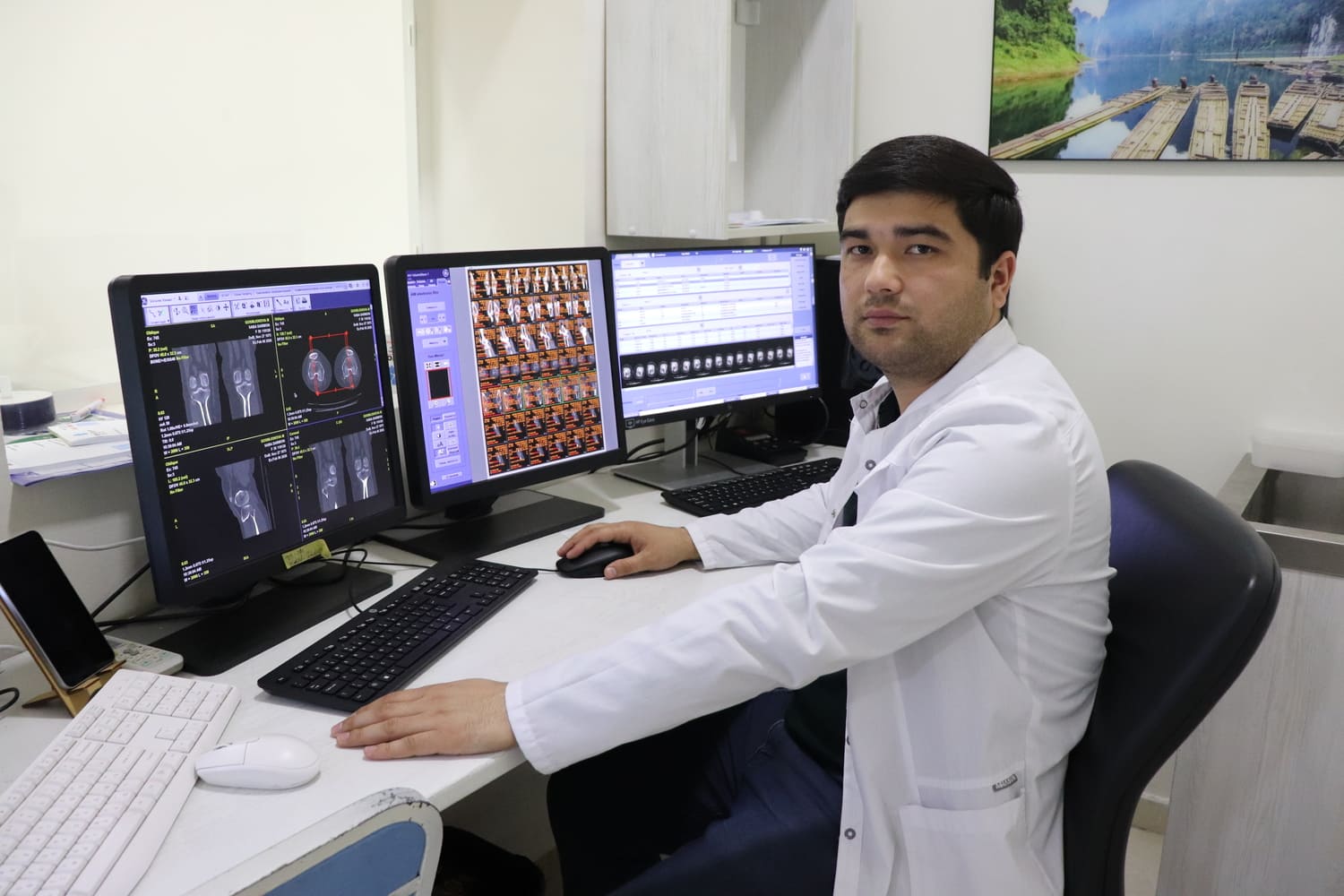
Ultrasound is the main and most accessible method: it shows the location, size and wall of the cyst and any "daughter cysts" inside it. It is ultrasound that defines the stage of the cyst. A CT scan (and MRI if needed) serves as a map before surgery: it shows how the cyst relates to blood vessels and bile ducts and whether there are cysts in other organs — read more in our CT scan article. A serological blood test detects antibodies to echinococcus and supports the diagnosis, but a negative result does not completely rule the disease out. To check the lungs, a chest X-ray or chest CT is done.
Put simply, cysts go through three kinds of stage: active (filled with fluid, sometimes containing daughter cysts), transitional (the inner membrane is starting to detach) and inactive (the contents have thickened or calcified). The choice of treatment depends largely on the stage.
Treatment options for liver echinococcosis
The surgeon chooses the method according to the number, size, location and stage of the cysts and the patient's general health. The main route is surgery: the cyst and its membrane are removed and the cavity is treated with special agents. A key point is to prevent the cyst contents from spilling into the abdomen.
| Point | Laparoscopic echinococcectomy | Open echinococcectomy |
|---|---|---|
| Access | Several small punctures in the abdominal wall; work through a camera | An incision under the ribs or along the midline |
| Who it suits more often | Conveniently placed, superficial, single or few cysts | Large, deep or multiple cysts, cysts close to bile ducts or major vessels, complicated cases |
| Recovery | Usually quicker, less pain, small scars | Usually longer, more days in hospital |
| Limitations | Not suitable for every cyst; the operation may be converted to open surgery if needed | Gives a wide view and full control in complex situations |
You can read more in our articles on laparoscopic and open echinococcectomy.
PAIR (puncture through the skin, aspiration of the fluid, injection of a special solution and re-aspiration) is a minimally invasive technique carried out under ultrasound guidance. It is used only in selected cases, for cysts at certain stages, and it is not suitable for everyone.
Albendazole is an anti-parasitic medicine. It is often prescribed in courses before and after surgery or PAIR, and for some small cysts it may be the only treatment. Because it can affect the liver and blood counts, blood tests need to be monitored; the dose and duration are decided only by the doctor. For very small, inactive (calcified) cysts, watchful monitoring is sometimes chosen instead.
Follow-up after treatment and recurrence
Echinococcosis can recur — a new cyst may appear or grow from remaining tissue. That is why regular ultrasound follow-up is carried out for several years after treatment: usually frequently in the first year, then once a year. Serology and CT are repeated if necessary. For preparation before an operation, see the pre-operative tests checklist.
How to prevent echinococcosis
- wash your hands with soap after contact with dogs, soil and livestock, and before eating;
- wash vegetables, herbs and fruit thoroughly under running water;
- drink only boiled or purified water;
- deworm dogs regularly, as advised by a vet;
- never feed dogs raw offal or liver; bury or destroy slaughter waste;
- do not let children kiss dogs or eat from the same dishes as them.
Another important liver topic is fatty liver disease. If a cyst has been found in your liver, do not delay seeing a surgeon — early assessment gives the widest choice of treatment and the lowest risk of complications.
Frequently asked questions
How do people get liver echinococcosis?
The parasite eggs pass out in dog faeces into soil, water and onto vegetables. People are infected by swallowing them with unwashed hands, vegetables or unboiled water; the disease does not spread from person to person.
Is a hydatid cyst of the liver dangerous?
Yes, especially a large one: it can become infected, rupture into the bile ducts or abdomen, or cause a severe allergic reaction. That is why it should not be left without medical follow-up.
Can echinococcosis be treated with tablets alone?
Albendazole can be the only treatment for some small cysts, but more often it is combined with surgery or PAIR. The doctor decides which approach fits based on ultrasound and CT findings.
Is laparoscopic surgery suitable for everyone?
No. It often suits conveniently placed superficial cysts; for large, deep or multiple cysts open surgery may be safer. The surgeon chooses the method.
Can echinococcosis come back after surgery?
Recurrence is possible, so ultrasound follow-up continues for several years. Following prevention measures lowers the risk of being infected again.
If my blood test is negative, does that rule out echinococcosis?
Not necessarily. Serology can be negative in some patients; the main diagnosis is based on ultrasound and CT images.
How can echinococcosis be prevented?
Wash hands with soap, wash vegetables thoroughly, drink boiled water, deworm dogs regularly and never feed them raw offal.
Related reading:
Doctors for consultation
Liver echinococcosis is treated by our surgeons using laparoscopic and open techniques. Our doctors in this field:




Book a consultation with a surgeon
Has an ultrasound found a cyst in your liver? A surgeon will review the images, the type of cyst and the right treatment.
Call now — +998 71 210-30-30Contacts
Address: 7A/3 Nurafshon street, Tashkent
Phone: +998 71 210-30-30 · WhatsApp: +998 95 662-30-30
Opening hours: Monday–Saturday, 09:00–18:00
E-mail: