


Useful articles

Breast Ultrasound or Mammography: Which One and When, and What BI-RADS Means
Doctor's blog
sbdrmn
Direction: Robotic surgery · All services
Breast ultrasound and mammography are the two main ways to examine the breast, but one does not replace the other. Ultrasound suits younger and pregnant women, while mammography is essential for screening after 40. Below, in plain language: how each test works, the best day to book, what a BI-RADS report means, and which signs mean you should see a doctor without delay.
Contents
How breast ultrasound and mammography work
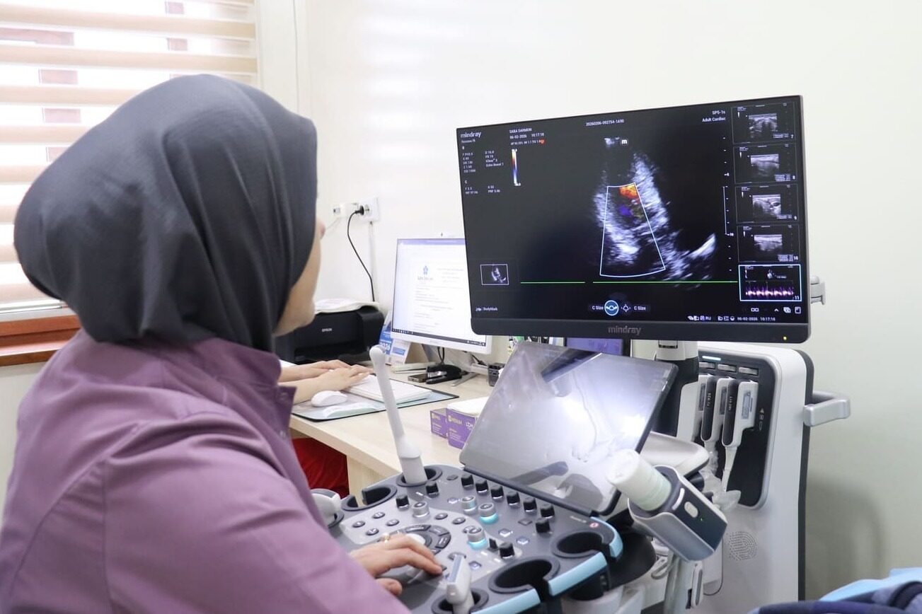
Both tests show what is happening inside the breast, but in different ways. Breast ultrasound uses high-frequency sound waves: a probe is placed on the skin with gel, and the reflected signal is turned into an image on the screen. There is no radiation, the test is painless and it usually takes 15–20 minutes. Mammography is a low-dose X-ray: the breast is compressed between two plates for a few seconds and imaged from two angles. The compression can be uncomfortable, but it is brief.
So the right question is not "which one is better" but "which one gives more information at my age and in my situation".
When ultrasound is the better choice
- Younger women (roughly under 35–40). Glandular tissue at this age is dense; on an X-ray it appears as a white background that can "hide" changes. For ultrasound, dense tissue is not an obstacle.
- Pregnancy and breastfeeding. Because there is no radiation, ultrasound is the first-line test; a lump, mastitis or a milk cyst (galactocele) is well seen on ultrasound.
- A lump has been found — cyst or solid? Ultrasound reliably tells a fluid-filled cyst from a solid mass. That alone answers many questions straight away.
- Dense breasts — as an addition to mammography.
- To guide a biopsy or drainage of a cyst.
When mammography is needed
Mammography is the main screening test (testing women without symptoms) from the age of 40. Its strength is detecting microcalcifications — tiny deposits of calcium. Sometimes they are the earliest sign of a tumour, and ultrasound often misses them. The screening interval (yearly or every two years) is set by the doctor according to your age, family history and previous results. Women whose mother or sister had breast cancer may be advised to start earlier.
The two tests complement each other
| Criterion | Breast ultrasound | Mammography |
|---|---|---|
| Method | Ultrasound, no radiation | Low-dose X-ray |
| Best suited to | Under 40, pregnant and breastfeeding women, dense tissue | Over 40, screening |
| Strength | Distinguishes cysts from solid lumps, sees armpit lymph nodes | Microcalcifications, an overall picture of the whole breast |
| Weakness | Often misses microcalcifications, depends on the operator's experience | Changes may be hidden in dense tissue |
| Discomfort | None | Brief compression |
In practice, a woman over 40 with dense breast tissue is often advised to have both: mammography gives the overall picture and ultrasound clarifies any suspicious area.
When to book: days 5–12 of the cycle
Women who menstruate are advised to have breast ultrasound on days 5–12 of the cycle, counting from the first day of the period. On these days the breasts are less swollen under hormonal influence, tenderness is lower and the image is clearer. Mammography is also easier to tolerate in the first half of the cycle. After the menopause the test can be done on any day. No special diet is needed, and the doctor will explain any ultrasound preparation. On the day of mammography do not apply deodorant or talc to the armpits — they can cast shadows on the image.
What a BI-RADS result means
The report often gives a BI-RADS category — an international scale showing how suspicious a finding is. It is not a diagnosis; it is a tool for choosing the next step.
| Category | Meaning | Usual next step |
|---|---|---|
| 0 | Not enough information | Additional imaging or comparison with previous images |
| 1 | No abnormality found | Routine check-ups |
| 2 | Clearly benign (simple cyst, stable fibroadenoma) | Routine check-ups |
| 3 | Probably benign, very low risk | Short-interval follow-up (often after 6 months) |
| 4 | Suspicious finding | Biopsy |
| 5 | Highly suspicious | Biopsy and specialist referral |
| 6 | Cancer already proven by biopsy | Treatment plan |
A "cyst" is a small fluid-filled sac; a simple cyst is almost always harmless and is usually just observed. A "fibroadenoma" is a common benign lump in younger women; if it grows or looks unusual, the doctor may suggest a biopsy or removal. Cyclical pain and lumpiness are covered in the article on breast pain and fibrocystic change.
When to see a doctor without delay
Do not wait for your next routine check if you notice: a new lump (especially one that does not go after your period); dimpling of the skin or an "orange-peel" texture; a nipple that has turned inwards; bloody discharge from the nipple; an enlarged lump in the armpit; a change in the shape or size of one breast.
Such signs often have a harmless cause, but only an examination gives a clear answer. Which test is needed and whether a biopsy is required is decided by the doctor after the examination and review of the images. Our ultrasound service and other departments are listed in the services catalogue.
Frequently asked questions
Which is better: breast ultrasound or mammography?
There is no universal "better". Under 40, in pregnancy and while breastfeeding ultrasound is usually chosen; over 40 mammography is the main screening test. Often the two complement each other.
On which day of the cycle should breast ultrasound be done?
On days 5–12, counting from the first day of the period. After the menopause it can be done on any day.
Is mammography dangerous?
The radiation dose is low, and from the age of 40 the benefit of screening is considered to outweigh the risk. In pregnancy ultrasound is usually done instead.
What does BI-RADS 3 mean?
It means "probably benign": the risk is very low, but the finding is watched for a while. A follow-up scan is usually arranged after a few months.
Can a fibroadenoma turn into cancer?
A simple fibroadenoma is a benign lump and as a rule does not become cancer. If it grows or changes, the doctor may suggest a biopsy or removal.
Can I have a breast ultrasound while breastfeeding?
Yes, ultrasound is safe during breastfeeding and is the first-line test. Feeding or expressing milk shortly before the scan makes the image clearer.
When is a biopsy needed?
Usually for BI-RADS 4 or 5, that is when a finding is suspicious. The decision is made by the doctor based on the images and the examination.
Doctors for consultation
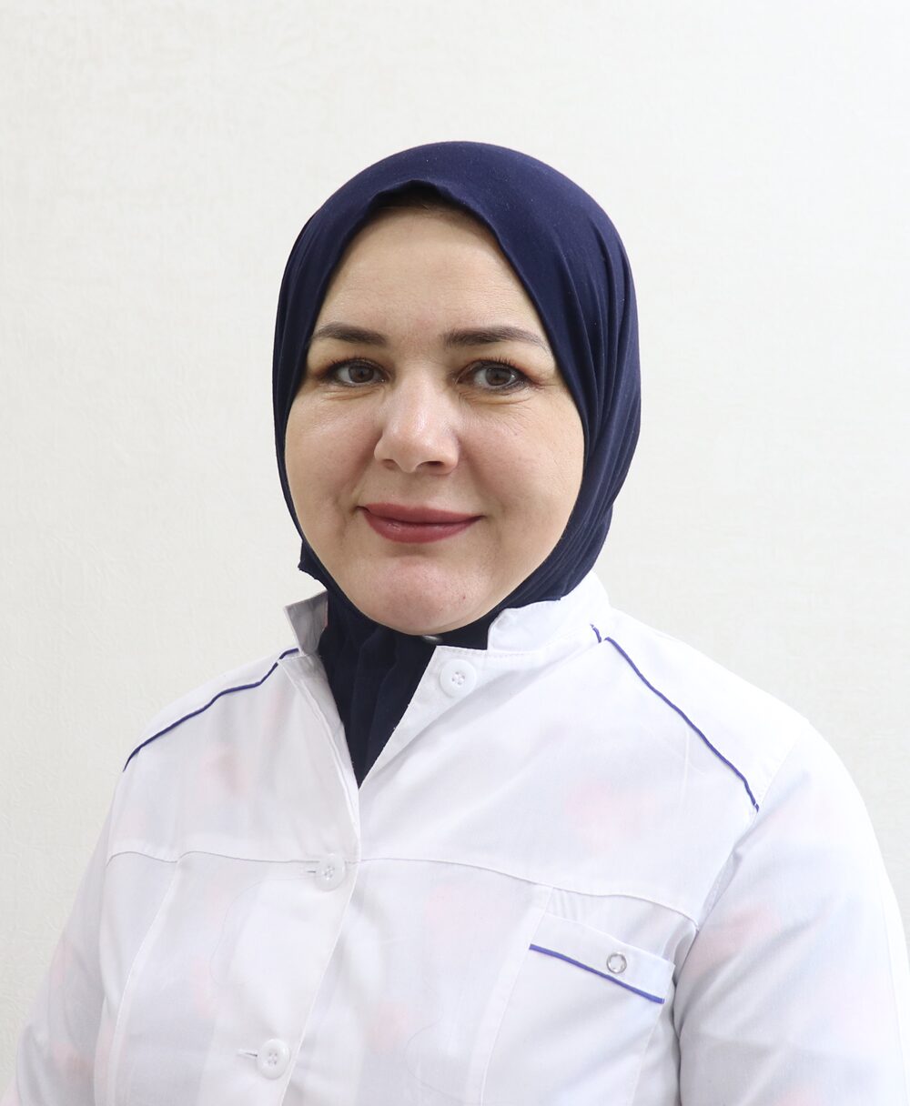
Breast ultrasound is performed by our ultrasound doctors. Our doctors in this field:



Book a breast ultrasound
Felt a lump or due for a routine check? Our ultrasound doctor will help you choose the right test.
Call now — +998 71 210-30-30Contacts
Address: 7A/3 Nurafshon street, Tashkent
Phone: +998 71 210-30-30 · WhatsApp: +998 95 662-30-30
Opening hours: Monday–Saturday, 09:00–18:00
E-mail: